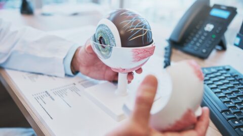
St. Antonius Hospital recently acquired a 3D lab. In the lab, the hospital develops 3D models that support medical specialists in preparing for operations. This allows them to perform operations more accurately and reduce the operation time. The 3D models can also help patients better understand their condition as well as treatment options and possible outcomes.
"In our 3D lab, we make so-called sight models and drill and saw templates," reports clinical physicist Vera Lagerburg of St Antonius Hospital. "A sight model is a 3D model of, for example, an organ, blood vessels or a joint. This can be used for patient explanations, education and training of medical staff and preparing for surgery. Drill and saw templates help the surgeon to place cuts and screws in the bone at exactly the right place. This is necessary when a bone needs to be straightened after a bone fracture or fusion."
The 3D model is based on a CT or MRI scan. Special software converts this scan into a visual model, which can then be 3D printed. The hospital uses fused deposit modelling (FDM), in which the 3D print is built up layer by layer from filament.
Get a good look at kidney from all sides
Lagerburg: "When a patient needs kidney surgery, for example, the surgeon can take a good look at the 3D kidney from all sides before the operation. This allows the surgeon to better prepare for the operation. And not only healthcare providers benefit; patients also better understand what is going to happen when they have a 3D model in their hands."
In the coming period, St Antonius Hospital will investigate for which conditions the use of the 3D lab provides the most added value. The focus will be on complex or rare conditions. More information is available here.
Supporting complex heart operations
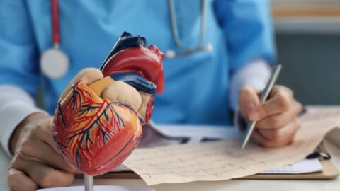
Other hospitals are also using 3D models to support the care they provide. One example is Catharina Hospital, which uses a 3D model to support complex heart surgery. The models help surgeons prepare for complex surgery and enable them to literally hold the heart in their hands.
Like St Antonius Hospital, Catharina Hospital also deploys the 3D model during discussions with patients, including to discuss the heart problem, possible treatment options and associated risks.
Precise localisation and removal of kidney tumours
Another example is the Princess Máxima Centre, which uses 3D models to help surgeons accurately locate and remove kidney tumours. The use of 3D models reduces the risks associated with kidney-sparing surgery, in which the healthy part of the kidney is preserved. The model can be viewed on a screen or as a hologram.
The Meander Medical Centre also uses 3D models. Last year, urologist Dr Anne Lont removed a kidney tumour using a 3D model. The patient had only one kidney, and a tumour had grown on this kidney that needed to be removed. The 3D model made it possible to remove the tumour whilst preserving the kidney, thereby ensuring the patient would not become dependent on dialysis.
Author: Wouter Hoeffnagel
Image: Darko Stojanovic via Pixabay