Researchers from the Academic Centre for Epileptology of Maastricht UMC+, TU Eindhoven and LUMC have succeeded in measuring activities still present in surgically removed brain tissue over a period of six days. The information gained could play an important role in the development of a so-called brain biopsy-on-a-chip.
In medical scientific research, the use of laboratory animals still plays an important role. However, according to the parties involved, society is increasingly critical of the usefulness and necessity of using laboratory animals for these purposes. In addition, it is increasingly clear that animal tests have only a limited predictive value for the outcome of follow-up studies in patients.
Animal-free models
For some time, therefore, researchers have been looking for alternative and non-animal models. For instance, researchers worldwide are working on manufacturing 'mini-organs' from human stem cells, also called 'organoids'. However, this method is not useful for all medical-scientific research. For instance, the human brain is so complex in structure and function that developing a mini-brain from human stem cells is still in its infancy.
The brain biopsy-on-a-chip could be a solution and an alternative. In this concept, doctors take a biopsy of brain tissue and make sure it remains active outside the body. Such a chip could be used for medical scientific research and reduce the use of laboratory animals for brain research.
Connecting brain tissue and electrodes
In practice, the concept consists of several layers. A circuit board with 120 electrodes (contact points) forms the basis here. On this circuit board, the researchers grow stem cells, which grow into a layer of nerve cells thanks to stimulation. These nerve cells must then make contact with the electrodes, which enables the measurement of electrical activity.
Researchers at the Academic Centre for Epileptology have succeeded in putting this concept into practice. Here, a piece of human brain tissue surgically removed during epilepsy treatment was placed directly on the nerve layer on the circuit board. Here, the nerve cell layer acts as a biological link between the brain tissue and the electrodes. The concept appears to work in practice; the model was kept in culture and showed epileptic activity after six days.
Evaluating personalised treatments
Maastricht brain researcher Dr Govert Hoogland praises the possibilities: "New experiments will have to show whether this is a useful model but our approach potentially provides a clinically relevant alternative for non-animal brain research of epilepsy, for example. It thus opens the way to evaluate new, personalised treatments."
The concept and culture media were developed by Dr J.P. Frimat and Dr R. Luttge, who are members of the Neuro-Nanoscale Engineering research group within the Microsystems division at TU/e. Dr J.P. Frimat now works in the Department of Human Genetics and Neurology (LUMC). Dr O. Schijns, Dr J. Dings, Dr K. Rijkers and Dr G. Hoogland, from the Department of Neurosurgery and the School for Mental Health and Neuroscience (MUMC+), applied the concept and culture media to human brain tissue. PhD student M. Hu and Prof. A. van Maagdenberg, from the Department of Human Genetics and Neurology (LUMC), analysed the measured activity. Further information about the research is available here.
Leading role in alternatives to animal testing
The Netherlands wants to play a leading role in the development of alternatives to research methods involving animal testing. In 2016, for instance, the Dutch government expressed its ambition to be an international leader in this field by 2025. Consequently, there are several developments in this field in the Netherlands.
One example is research by neurobiologist Nael Nadif Kasri of Radboudumc. In a multiple publication, Kasri illustrates the potential of so-called 'mini-brains' grown from skin cells. These show a characteristic pattern of electrical discharges. Research shows that this pattern is different in people with neurological disorders or intellectual disabilities.
Basic rhythm
Kasri put cultured brain cells into small trays, where the cells make contact with each other. This creates a network of interconnected brain cells, which send electrical signals. The operation here resembles the brain cells in the human head. After some time, the cultured brain cells give off a certain pattern. This creates a kind of basic electrical rhythm, which can be picked up using a chip in the tray.
For anyone with healthy brain cells, this basic rhythm is the same. Cultured brain cells from someone with a brain disorder, for example, also show a basic rhythm, but this rhythm differs from the 'normal' basic rhythm. For example, people with MELAS (Mitochondrial Encephalomyopathy, Lactate Acidosis and Stroke) appear to show the same abnormal pattern, which is characteristic of their condition.
SMART Organ-on-Chip
Another example is the Dutch consortium hDMT, which brings together 10 research groups from eight universities. The parties work together with companies, knowledge institutions and foundations. A total of 33 parties are involved. Together, they want to develop a standardised and modular platform for organs-on-chips called SMART Organ-on-Chip.
Organ-on-Chip Development Centre
More recently - in May 2023 - the Netherlands Organisation for Scientific Research (NWO) invested more than three million euros in the Organ-on-Chip Development Centre, led by Dr Andries van der Meer. The centre is working on setting up a facility called hDMT INFRA OoCDev. This facility should enable the development of innovative organs-on-chips by pooling frontier infrastructure and technical expertise.
"This facility will make it possible to develop organs-on-chips that are truly next generation, with working three-dimensional tissues, blood flow and dynamic loading," explains Dr Van der Meer. "The Organ-on-Chip Development Centre provides a central location for this pioneering work, not only for local researchers, but precisely also for all scientists from other institutes affiliated to the national 'Institute for human Organ and Disease Model Technologies (hDMT)'."
Author: Wouter Hoeffnagel
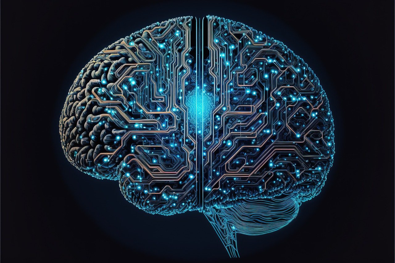
Photo: Kohji Asakawa via Pixabay