A new method makes it more predictable whether a weakness in the abdominal artery (aorta) is dangerous. The technique can help treat patients earlier and more targeted, and make life with an aneurysm less stressful.
Specifically, the method helps assess an aneurysm in the aorta, which is a dilation in a blood vessel. In many cases, an aneurysm does not cause any symptoms. However, does the blood vessel rupture? If so, this can be life-threatening.
Assessing risk better
In many cases, an aneurysm is discovered by chance. In that case, checks follow, looking to see if the dilation is widening. "Doctors now mainly look at the growth and diameter of the aorta," explains Catharina Hospital researcher Floor Fasen. Fasen developed the method. "But sometimes a small aneurysm does rupture, and a large one does not. So maybe there are better ways to assess risk."
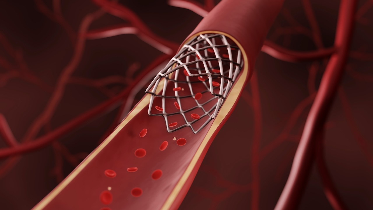
The method developed by Fasen allows the wall of the aorta to be viewed from the inside. The method has been used during EVAR procedures at Catharina Hospital. This involves placing a stent on the weakened spot in the aorta through a small opening in the groin. The stent traps the pressure of the blood, sparing the fragile vessel wall.
Measuring aortic wall thickness
Phases inserted a thin tube into patients during EVAR procedures. A small ultrasound head, similar to a pregnancy ultrasound, was placed at the end of this tube. "This allowed me to measure the wall thickness of the aorta," says Fasen. "This cannot be done with an ultrasound through the top of the abdomen, as my colleague Esther Maas did. But my method was more invasive, so only suitable for a small group of patients: people who were undergoing EVAR surgery anyway."
Through the measurements, Fasen collected a lot of data. The researcher uses this for analyses and the development of an AI model that automatically recognises features in the aortic wall. "What this model does now is measure wall thickness. Then we try to make connections between wall thickness, calcifications and blood clots," says the PhD student. "Those calcifications are hard pieces in the otherwise flexible vessel, and cause the wall to move less well. We think that can increase the risk of rupture."
Practised on porcine aortas
To train the model, Fasen practised in a laboratory with pig aortas, which are very similar to human aortas. "I built a kind of fake belly made of gelatine, into which I placed the pig aortas," reports Fasen. "This creates a realistic environment for the ultrasound, similar to the situation in the human body."
However, the AI model that Fasen is working on is not yet able to predict for each patient whether an aneurysm will rupture. However, the AI model does make patterns visible and provides insight into the growth of aneurysms and changes in the vessel wall. Fasen: "In the future, you obviously hope to be able to say something for each patient individually, but for the time being this invasive method proves difficult. Still, what we have learned now is valuable. A subsequent researcher can build on the insights we now have into how wall thickness, calcifications and clots are related in aneurysm growth."
Ticking time bomb
Fasen hopes that thanks to her development, doctors will be able to better predict in the future whether an aneurysm puts someone at real risk. This could not only save unnecessary scans and hospital visits, but also relieve patients. For example, many patients feel like they are walking around with a ticking time bomb. "Especially when you know it can rupture, but don't know when or if it will happen," says the PhD student.
"I studied electrical engineering prior to my PhD research, so my background is mainly technical," concludes Fasen. "When I started this research, I had yet to learn everything about blood vessels. But I really wanted to do something for healthcare. Now I am sure I want to continue working in the medical-technical world."