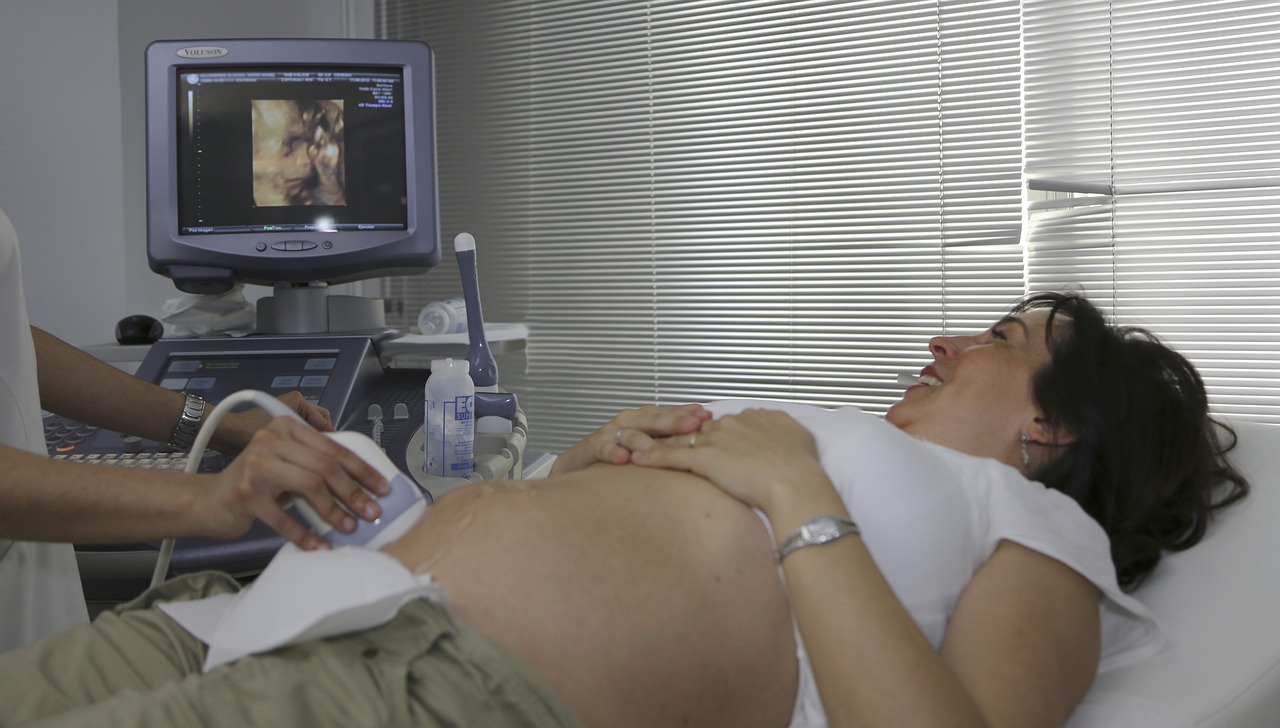

Researchers from TU/e, Catharina Hospital and Philips have succeeded in improving the image quality of ultrasound by using two echo heads. By taking ultrasound images with multiple heads simultaneously and then combining these images, the image quality of an ultrasound can be significantly improved.
Ultrasound uses ultrasonic sound waves to look inside a patient's body. It is a well-known and widely used technique, used in a variety of healthcare specialities. However, ultrasound also has disadvantages. For instance, you depend on the ultrasound technician to take exactly the right images. Because sound waves collide and bounce off different tissue in the body, muscle and fat tissue, among others, can scatter the waves and produce noise on the images.
Many times cheaper than alternatives
In some cases, this makes it necessary to use alternative techniques. A considerable disadvantage, as an ultrasound is usually many times cheaper. By comparison, an ultrasound often costs sixty to eighty euros each, while the cost of a CT or MRI scan is easily a thousand euros.
Researchers at Eindhoven University of Technology (TU/e) are therefore investigating ways to improve ultrasound quality. PhD student Vera van Hal did her PhD on whether you can make an ultrasound with two heads at the same time, which is also called multi-aperture ultrasound. She is the first to succeed in making such an ultrasound. Van Hal conducted experiments using pig aortas in a set-up that mimics the body as well as echoes on volunteer subjects at Catharina Hospital.
Sending out sound waves twice also produces noise twice
"For that, I did have to solve some problems," Van Hal explains. "Because sending out sound waves twice also means receiving two echoes, and twice noise and signals that can interfere with each other. Previous PhD students didn't quite figure that out."
"With multi-aperture ultrasound, we use principles from radar technology, alternating transmission and then listening with two heads at the same time. Each head transmits one signal and captures two: the echo of its own signal and that of the head next to it. So with two heads, that yields four signals. And then we combine all four signals coming back in using smart software."
Combining signals into one ultrasound image
Van Hal managed to link and combine the four signals in time and location into a single ultrasound image on the screen. "This way, we were finally able to solve a big puzzle that researchers had been working on for a while. That's really tremendously cool."
The results are promising. For example, combining the four signals produces a much sharper and more detailed image. In addition, structures that were previously not visible on an ultrasound image can be made visible by looking from a different direction.
"That has everything to do with the fact that we can only capture the echoes that are reflected back to the echo head," Van Hal explains. "But if we combine the images, then we see AND less noise and we can fill the 'holes' in the image from one head with image from the other head. So we see more information in the full ultrasound image."
Aneurysm monitoring
In her research, Van Hal collaborated with Judith Fonken, who received her PhD from the Faculty of Biomedical Engineering on 25 March on modelling an aneurysm of the abdominal aorta. Central to her research was being able to predict the development of an aneurysm based on reliable measurement data.
Van Hal explains: "An aneurysm of the aorta is a dangerous dilation of the vessel wall of the largest artery in our body. Such an aneurysm is often discovered by chance and then watched closely. Because when it ruptures, the internal bleeding is so severe that it is life-threatening." Therefore, the dilation in the aorta is closely monitored, and surgery is performed if the diameter exceeds the limit of 55 mm in men and 50 mm in women.
Increasingly questioned need
Such an operation is a major operation on the part of the patient. Increasingly, however, the need for the operations is being questioned. For instance, it is suspected that even large aneurysms can be safe, while smaller ones sometimes rupture anyway. The cause of this is unknown.
"With our double-headed echo, the field of view, contrast and resolution improves so much that we can clearly see the entire circumference of the aneurysm, as well as the surrounding tissues," says Van Hal.
Determining vessel wall stretch
Thanks to the extra information and better image quality provided by the double-headed ultrasound, it is possible to determine the movement and stretch of the vessel wall, among other things. Van Hal: "And not only can we measure this well, we can also show it with colours over the moving images. When you then compare the images of a single head with those of a double head, you immediately see that with the latter, the images are much more accurate and contain less noise."
Among other things, the better image quality of a double-headed ultrasound allows local differences in strain in the material to be properly analysed. Thus, a picture of the difference in material properties in the aneurysm can be obtained. Important, because strain indicates forces and tension. If it rises too high, it destroys the vessel wall. "With our new ultrasound techniques, we can accurately determine this stretch not only in the vessel wall, but also in the material stuck inside against the vessel wall - which is called the thrombus. Since we don't really know how this affects the likelihood of the dilation rupturing, this could start to provide a lot of important information."
Van Hal concludes, "This saves healthcare money and people an unnecessary operation. It may also mean that people for whom it is necessary are actually helped earlier. In this way, our research will hopefully deliver real improvements for everyone in the future: patients, doctors and society."
More information is available here.
Author: Wouter Hoeffnagel
Image: MedicalPrudens via Pixabay